
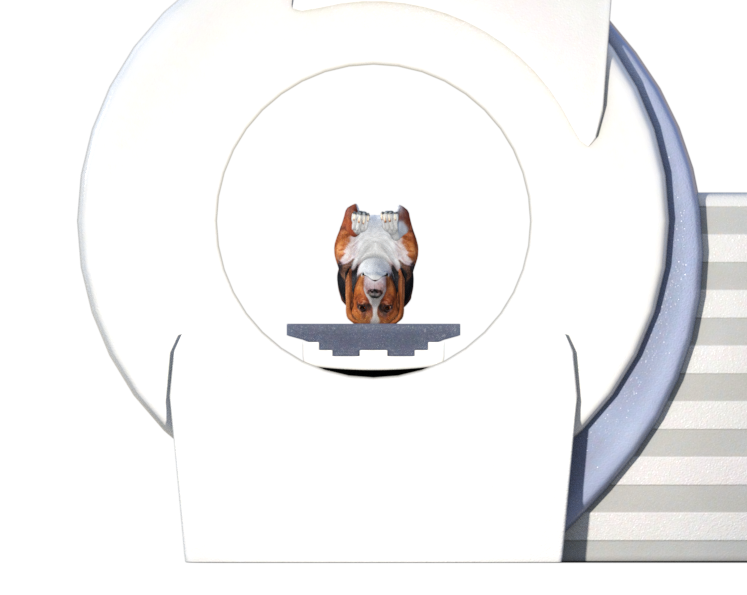
Positioning for a brain MRI
- Animal on its back
- Neck straight
- Mandible should be parallel to the plane of the table top
Please note: instructions and diagram are meant to convey basic positioning in the simplest way possible. We do not show pads or straps and it is assumed, for example, that the spine of the dog below would be supported. If your anesthesia or coil setup does not allow for head-in positioning, simply rotate the animal 180 degrees.
Do you have a question about positioning or a suggestion to improve our 3D illustrations? We welcome your input! Please comment below.


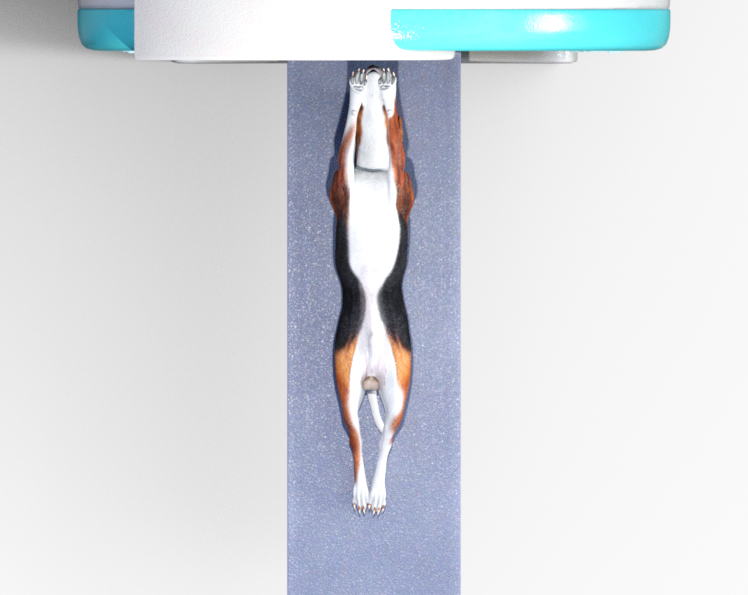
3D Interactive model
Controls: Left mouse button rotates, Right mouse button pans, wheel zooms.
Example #1
There are several different variations and coil placements used in the field. The version below was generously submitted to us by Ken Glatt of Companion MRI. In this case, the brain of a Cavalier King Charles Spaniel is imaged with a GP Flex coil.

Example #2
There are several different variations and coil placements used in the field. The version below was generously submitted to us by Ken Glatt of Companion MRI. In this case, the brain of a Doberman is imaged with a Quad Knee Coil.


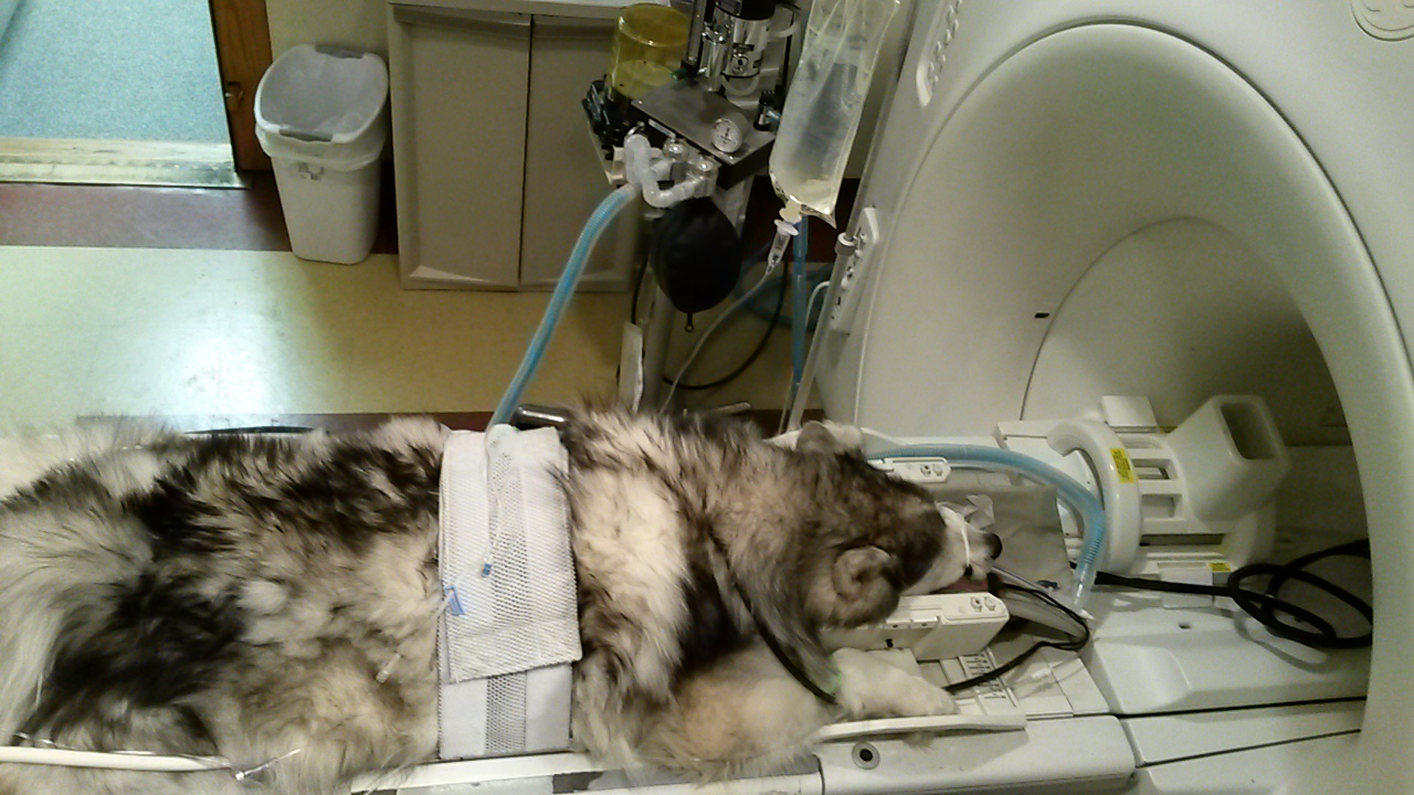
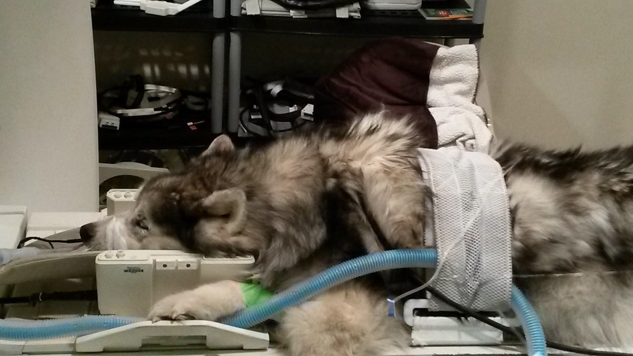
Example #3
There are several different variations and coil placements used in the field. The version below was generously submitted to us by Ken Glatt of Companion MRI. In this case, the brain of a Siberian Husky is imaged in the ventral position with a Quad Knee Coil.
0 Comments